Hyperpigmentation after sclerotherapy: a matter of choices, not just bad luck
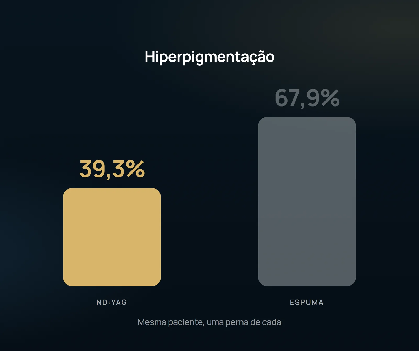
In the Swiss trial that compared polydocanol foam and Nd:YAG in the same patient, hyperpigmentation was 67.9% on the foam side against 39.3% on the laser side. What hardly anyone mentions is the explanation of the authors themselves — and the three levers that she puts in the hand of those who mount the session.
 View the video lesson
Post-sclerotherapy hyperpigmentation is usually treated as an accident — something that depends on the patient's skin. The essay by Parlar et al. deals with the subject in another way: the authors explain the fee itself, and the explanation transfers responsibility to those who set up the session.
What was measured
Fifty-six women with primary leg telangiectasis and reticular veins (C1A) were randomized to receive polydocanol foam at 0.5% on one leg and Nd:YAG laser at a long pulse of 1064 nm — two sessions at 6 weeks apart, followed at 6 months. The telangiectasis were 0.2 to 1 mm and the reticularis 1 to 2.9 mm, in an area marked 10 x 15 cm on each side. Age from 23 to 66, mean 46. Fifty-one of the 56 completed the study.

The phrase that changes the conduct
In the discussion, the authors wrote that the relatively high pigmentation rate observed is due to the fact that they chose polydocanol in foam. The mechanism is the same that gives effectiveness to foam: the foam agent replaces the blood present in the vessel, comes into full contact with the endothelium and adheres better to the wall, while the liquid solution mixes with the blood and loses effect by dilution. Efficacy and pigmentation come through the same door.
The three levers
The first is the form: because of this side effect, some experts prefer liquid sclerotherapy rather than foam for leg telangiectasia, with the same concentration of polydocanol at 0.5%. The second is the concentration: the authors raise that 0.25% instead of 0.5% to produce foam could be potentially sufficient, causing less pigmentation — explicit hypothesis of them, not outcome measured in this trial. The third is compression: there is evidence that the use of compression stocking for 3 weeks after sclerotherapy improves vessel clearance and reduces pigmentation rate by decreasing thrombi formation and minimizing inflammation and angiogenesis. In the trial, the sock was class II Swiss, from 26 to 32 mmHg, used during the day.
The conversation before the session
The authors are explicit: it is important to explain to the patient the increased risk of pigmentation problems related to foam sclerotherapy. And pigment is not the only side effect in play: matting appeared equally frequently in both therapies, with a prevalence of 20%, and there was limited skin necrosis in one patient in each group. The baseline literature records, for sclerotherapy, hyperpigmentation in up to 60% of cases, matting in 9.2%, necrosis in 3.5% and allergic reaction in 0.6%.
What the laser charges in return
There was a statistically significant difference in pain perception between therapies (P = 0.003): laser was felt as more painful, even with fluence of 100 to 200 J/cm2, spot of 3 to 7 mm, pulse of 10 to 50 ms and a cold air system cooling the skin. Nevertheless, patient satisfaction at the last visit was the same on both sides (median 5) and subjective perception of improvement did not differ (P = 0.58). The same woman, with the stain on one side and the pain on the other, did not separate the two treatments.
Therefore, the authors' conclusion is not to score, but rather to indicate: the Nd:YAG of 1064 nm is suitable especially in case of needle phobia, allergy to sclerosing and in the presence of small vessels with telangiectasic matting — including vessels smaller than the diameter of a 30-gauge needle — while sclerotherapy also treats the feeding vein in the same session. And there is no option that does not pigment: the most observed complication with long pulse Nd:YAG in the literature is hyperpigmentation, with frequency of 16 to 62% according to the study.
What the study does not authorize to state
The inclusion criterion limits the transfer of numbers: only phototypes of Fitzpatrick 1 to 3 and only women entered. Higher phototypes have not been tested in this assay, and the pigmentation rates described here cannot be extended to them. The trial is open, and side effects were also checked by the treating physician, who was not blinded to treatment — agreement with blind evaluators was only moderate in laser (ICC 0.53) and strong in sclerotherapy (ICC 0.73). The follow-up was 6 months, with 5 losses. And there is a statistical point that usually passes beaten: the sample size was calculated to detect a large difference — clearance of up to 20% in laser versus at least 45% in sclerotherapy (α = 0.05, β = 0.20). Finding no difference in such a test is not the same as demonstrating equivalence; this is not a non-inferiority study. The one who decides the conduct of each leg is the evaluation made by a vascular surgeon.

Content addressed exclusively to health professionals. No individual result is guaranteed: the indication depends on clinical evaluation and imaging examination of each patient (CFM 2,336/2023).